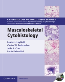
Image for Musculoskeletal Cytohistology

Musculoskeletal Cytohistology

Part of the Cytohistology of Small Tissue Samples series
See all formats and editions

Each volume in this richly illustrated series, sponsored by the Papanicolaou Society of Cytopathology, provides an organ-based approach to the cytological and histological diagnosis of small tissue samples.

Benign, pre-malignant and malignant entities are presented in a well-organized and standardized format, with high-resolution color photomicrographs, tables, tabulated specific morphologic criteria and appropriate ancillary testing algorithms.

Example vignettes allow the reader to assimilate the diagnostic principles in a case-based format.

This volume describes the cytologic, small core biopsy, immunohistochemical and molecular diagnostic features of musculoskeletal lesions.

It provides the cytopathologist and histopathologist with a comprehensive survey of diagnostic approaches and diagnostic features for benign and malignant lesions.

Each chapter contains a detailed summary of differential diagnostic features, supported by high-quality images and case studies.

The print edition includes a CD-ROM offering all images in a downloadable format.

With over 500 photomicrographs, this is an important resource for practising pathologists and residents in pathology.

Read More
Special order line: only available to educational & business accounts. Sign In
Product Details
Cambridge University Press
1139209949 / 9781139209946
eBook (EPUB)
23/02/2012
English
801 pages
Copy: 10%; print: 10%